

Living Networks offers custom-tailored high-throughput drug screening services based on our advanced vascularized human tissue model.
We support your research with scalable, physiologically relevant models designed to accelerate insight and maximise translational success.

Pro-angiogenic drug screens
in various environments
This may include diabetic tissue or skin models to assess therapies promoting vascular growth.

Endothelial cell toxicity screening
Detecting off-target drug effects on vascular health.

in cancer microenvironments
Identifying compounds that inhibit tumor angiogenesis and/or remodel tumor associated vasculature.

Fully automated software
Flexible drug regimen design.
Including combinatorial screens of combination therapies.
Seamless compatibility
Our models are compatible with standard multi-well plate formats for streamlined integration into existing workflows.
Anti-angiogenic drug screens
for morphometric measurements
Compatible with high-content 3D confocal fluorescence imaging, eliminating bias and human errors.
Services

Our platform offers a wide array of applications, enabling researchers and developers across disciplines to accelerate preclinical development.
Applications


Biotech & Pharma Companies
We offer DIY cell-culture kits and a dedicated fully automated angiogenesis image-analysis software, for laboratories striving to improve precision, throughput and automation of angiogenesis assays in various 3D microenvironments.

Oncological Centers
For outsourcing preclinical development. Our screens are complementary to standard in vivo models (e.g. PDX model, Matrigel plug assay, CAM assay), while delivering more precise information on the specific role of the human microvasculature. Evaluated at ultra-high throughput.
Cosmetics and Research & Development laboratories focusing on couperese or vascular skin applications and other indications that involve angiogenesis and vascular remodelling in the skin.
Laboratories
Researching the impact of tumor microenvironment on drug efficiency. Our assays are capable of correlating tumor growth with vascular remodelling under various drug regimens including combination therapies. We also offer development of custom. models, such as vascularized immune tumor microenvironments.

Cosmetics R&D laboratories
Process outline
Step 1. Delivery
You deliver compounds to be tested.


Step 2. Modelling
In collaboration with you, our experts establish an advanced 3D human tissue model to optimally meet your needs.

Step 3. Screening
Using the established model, we perform in-house screening services including dose-response studies and/or combinatorial tests.
Step 4. Analysis
Analysis of the results and delivery of a detailed quantitative report including summary of the most important findings.
Case studies
Explore our selection of case studies to see how Living Networks’ vascularized human tissue models are applied in real-world research. From angiogenesis assays to cancer microvasculature imaging, these examples highlight the precision and versatility of our platform in addressing diverse questions in drug discovery and development. Each case study provides a closer look at how our technology generates reproducible, physiologically relevant insights that help accelerate innovation.
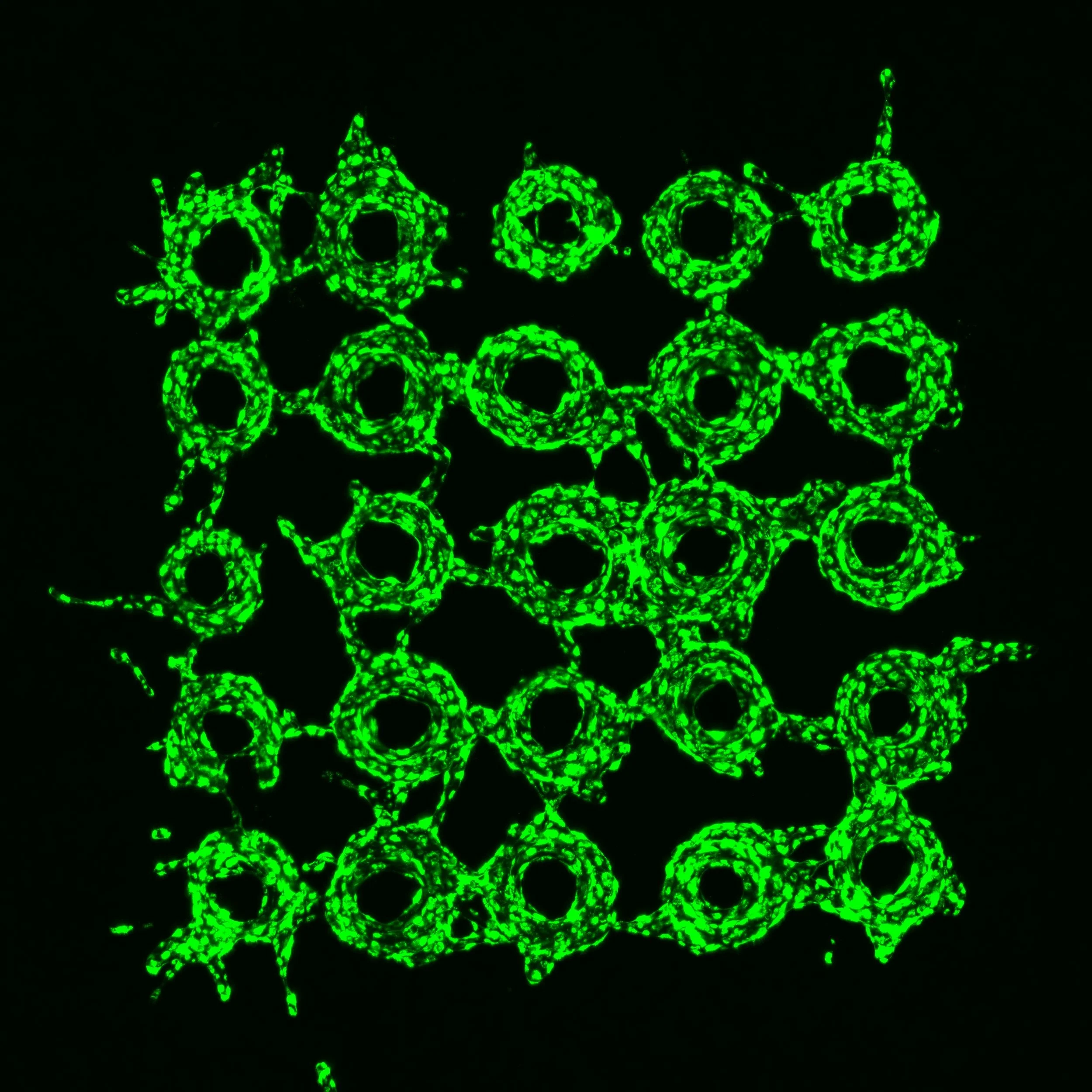
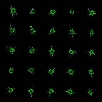
Case study I
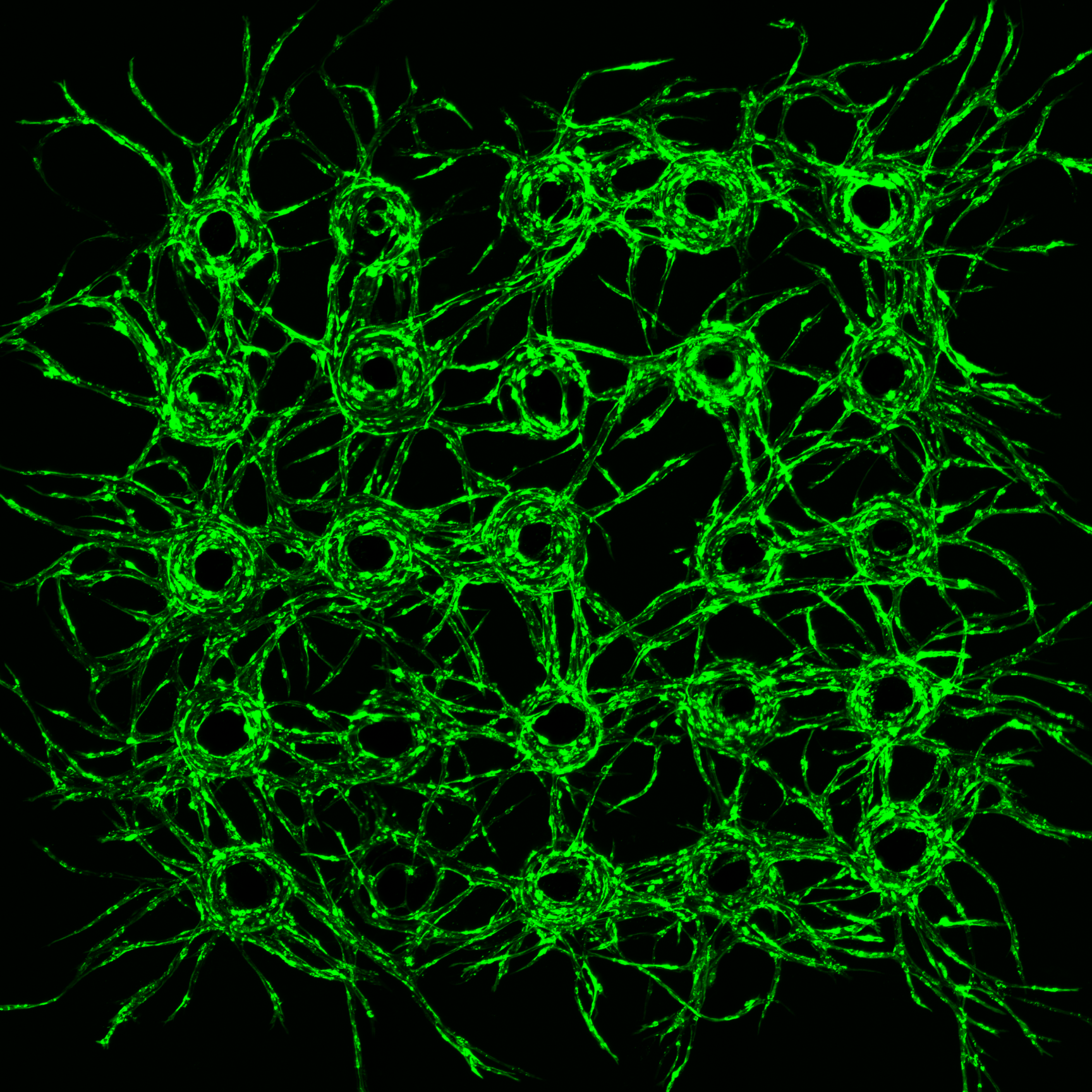
VTμA angiogenesis sprouting assays (bead spacing 1500 μm).
Confocal images show growing vascular microtissues visualized with GFP (green).
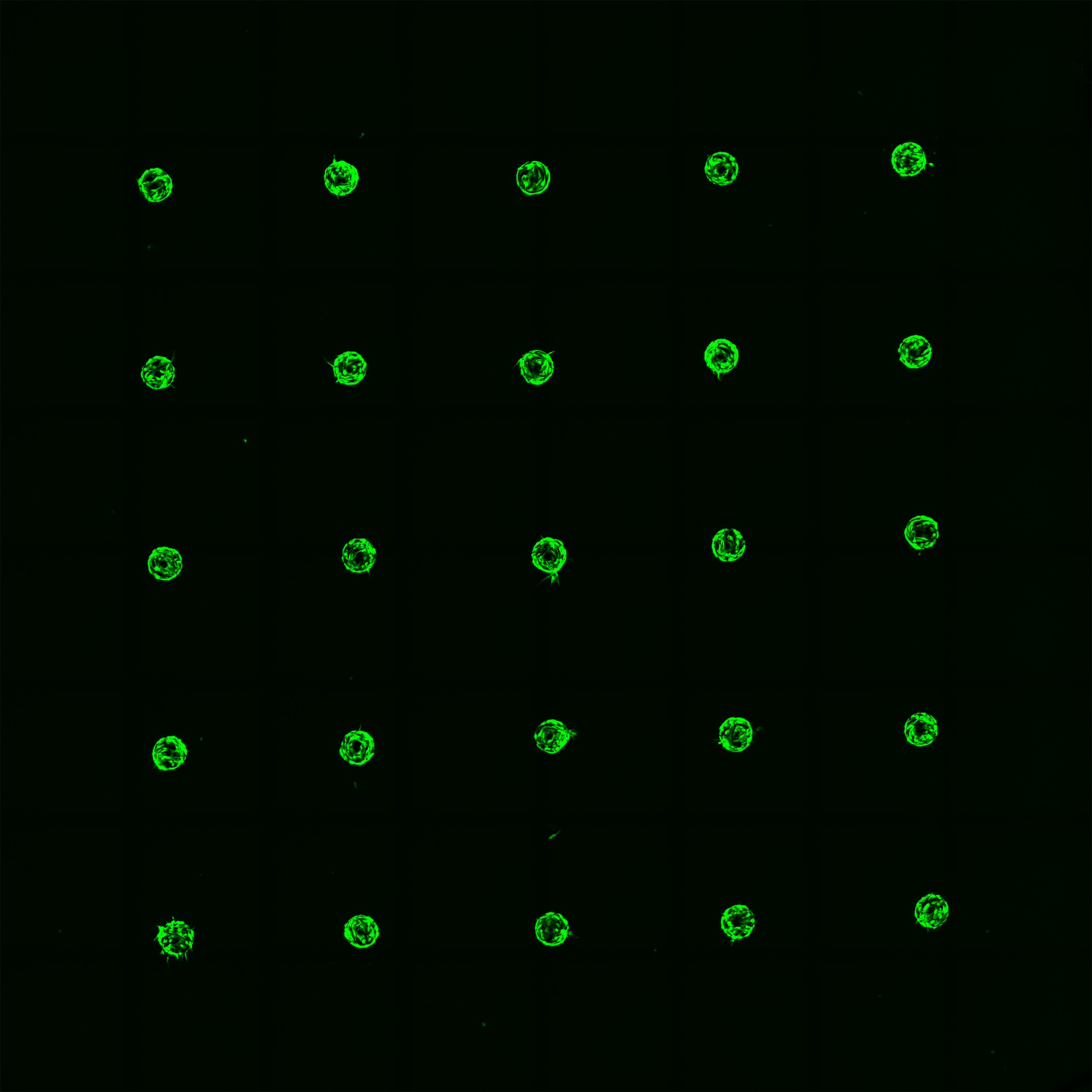
Day 1

Day 5
Case study II
VTμA: vasculature disintegration assays (bead spacing 500 μm).
VTµA of cervical cancer microenviroment.
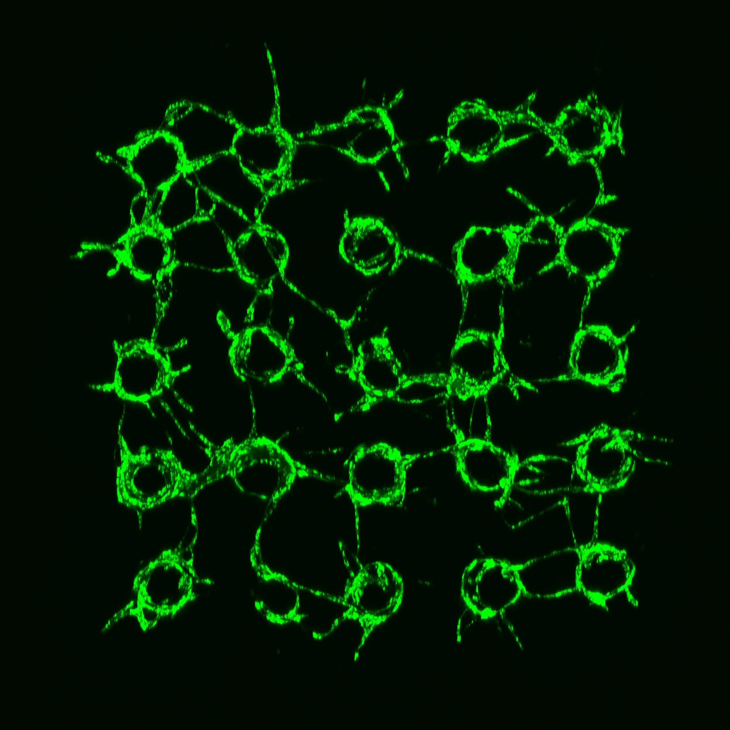
VTµA of breast cancer microenviroment treated with 100 nM Axitinib for 48h.
Case study III
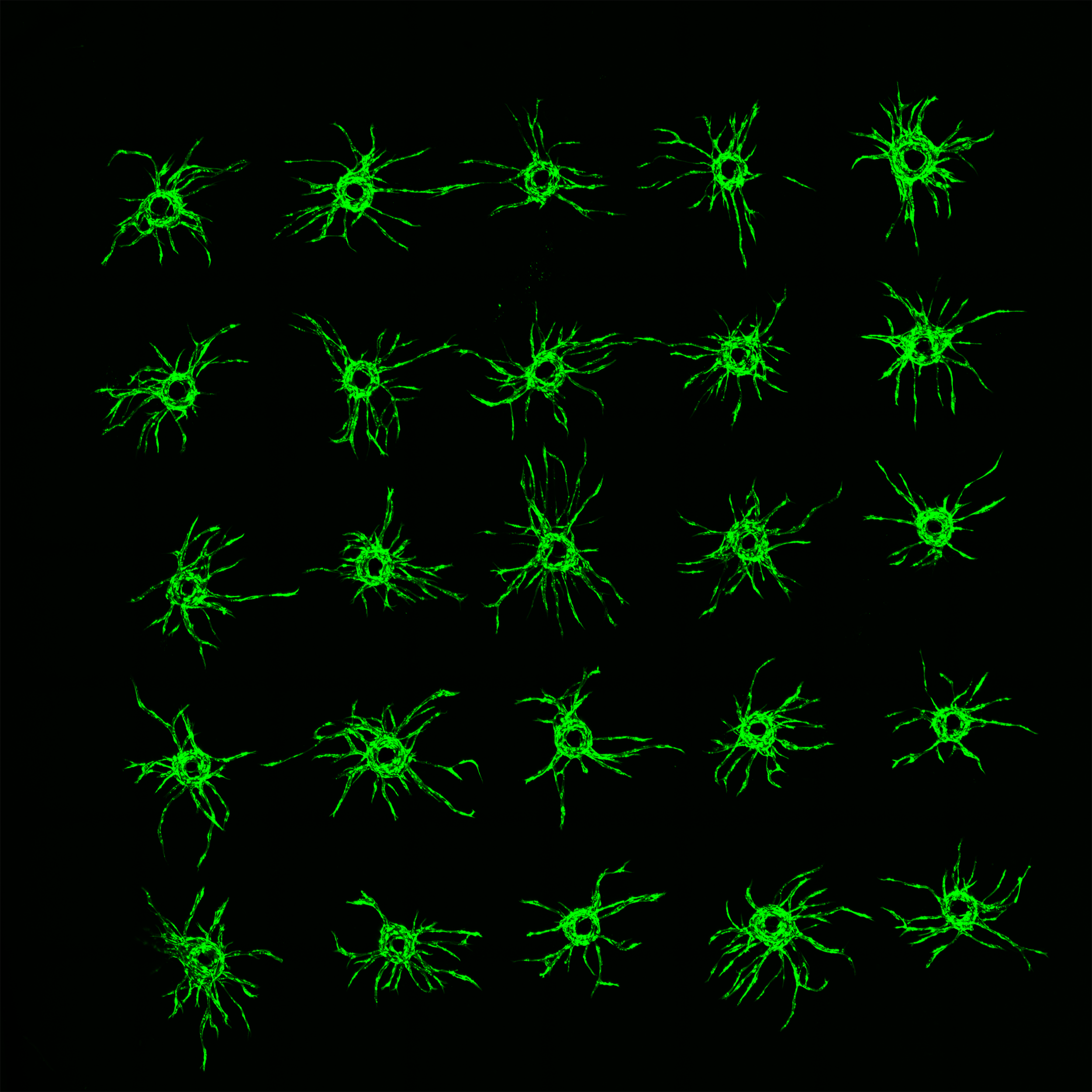
Morphology of the cancer microvasculature depends on the type of cancer.
Vascular microtissues visualised with GFP (green). Cancer cells not labeled.
Direct time-lapse imaging of cancer-cell migration and intravasation.
Microvasculature is visuallized with RFP (red) and cervical cancer cells with GFP (green).
Day 7
VTµA of breast cancer microenviroment treated with 1000 nM Taxol for 48h.
Pic 2. Breast cancer microenvironment (MDA-MB-231-Br),


Pic 3. Cervical cancer microenvironment (HeLa);


Case Study IV

Pic 1. Breast cancer microenvironment (MCF-7)


